


Posted by: Ophthalmology Associates of the Valley in Eye Health

Keratoconus develops slowly, and by the time it causes noticeable blur, the cornea has already been thinning and reshaping itself for months or years.
Glasses and even standard soft contacts often can’t keep up with the changes, and many patients bounce between prescriptions without ever landing on one that works long-term.
Corneal cross-linking treatment works by strengthening the fibers that give the cornea its shape. For patients with a documented pattern of progression, stabilizing the cornea is often the first step to finding a better vision correction solution.
Keep reading to learn more about corneal cross-linking and how it can help treat keratoconus.
What Keratoconus Does to the Cornea
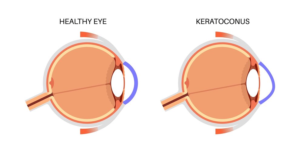
The cornea is normally shaped like a dome that’s evenly curved so light focuses directly onto the retina. In those who have keratoconus, the collagen fibers that hold that dome in place lose some of their natural bonds, and the tissue gradually thins in one area. As it thins, that area bulges outward, causing the cornea to be more of a cone shape rather than a sphere. This causes irregular astigmatism that changes rapidly and leads to glare, ghosting, and blurred vision that contact lenses fitted for a normal corneal curve cannot correct.
Keratoconus most often begins during the teenage years or early twenties, when the cornea is still undergoing its own natural growth, and it tends to level off with age.
Patients who rub their eyes frequently, wear poorly fitted lenses, or have a family history of the condition face a higher likelihood of both developing it and of it progressing quickly, a pattern that eye doctors track closely once other corneal diseases and disorders have been ruled out during the diagnostic workup.
How Does Corneal Cross-Linking Work?

Corneal cross-linking targets the weakness that keratoconus creates at a structural level.
During the procedure, an eye doctor applies riboflavin, which is vitamin B2, directly onto the surface of the cornea in repeated drops over roughly half an hour. Once the tissue has absorbed the riboflavin, it’s exposed to a controlled dose of ultraviolet A light. The combination triggers a reaction that forms new bonds between the collagen fibers, adding connective links between strands of tissue that had drifted apart as the cornea thinned. Those new bonds increase the rigidity of the cornea without altering its shape or size.
Cross-linking can be performed with the corneal epithelium on or off. The corneal epithelium is the eye’s thin outer layer. It can either be left intact or temporarily removed. Removing the epithelium, known as epi-off, allows the riboflavin to soak into the deeper stroma faster and more evenly, which is why it remains the more established approach for achieving consistent strength. Leaving the epithelium in place, known as epi-on, shortens the recovery window and reduces initial discomfort, but the riboflavin has a harder time penetrating to the same depth.
An eye doctor considers corneal thickness, the degree of scarring, and the extent of keratoconus progression before recommending one method over another.
What Cross Linking Can and Cannot Reverse
Cross-linking is a stabilizing treatment. It halts the progressive steepening that comes from ongoing keratoconus, but it does not flatten a cornea that has already taken on a cone shape, and it will not sharpen vision on its own the way glasses or a refractive procedure would.
However, by stopping the disease from advancing further, cross-linking reduces the odds that a patient will eventually need a corneal transplant, a possibility that becomes more likely once keratoconus reaches advanced stages.
Some patients later become candidates for Intacs corneal implants, tiny plastic segments placed within the cornea to flatten its shape and improve how contact lenses or glasses fit once the underlying disease has been stabilized.
Who is a Candidate for Corneal Cross-Linking?
At Ophthalmology Associates of the Valley, the oldest established multispecialty ophthalmology group in the San Fernando Valley, eye doctors look for documented progression before recommending cross-linking, meaning corneal topography scans taken months apart show measurable steepening, thinning, or a shift in irregular astigmatism that glasses and standard lenses can no longer keep up with.
Younger patients are watched especially closely, since keratoconus tends to move fastest in the teens and twenties and slows considerably by the thirties and forties. They will also look at your corneal thickness, since cross-linking requires enough tissue depth to safely absorb the riboflavin and tolerate the UV-A exposure without risking damage to deeper structures of the eye.
Patients with post-LASIK ectasia or pellucid marginal degeneration, both of which involve similar corneal weakening, may qualify for the same treatment.
A comprehensive eye exam, including topography and corneal thickness measurements, is the only reliable way to confirm whether cross-linking is the right next step or whether continued monitoring makes more sense for now.
What is Treatment and the Recovery Like?

Cross-linking is typically performed as an outpatient procedure that takes under two hours from start to finish, most of which is the riboflavin saturation period rather than the UV-A exposure itself.
First, your eye doctor will instill numbing drops to keep the eye comfortable throughout, and patients go home the same day with a bandage contact lens protecting the surface while it heals.
The first several days after epi-off treatment may be uncomfortable, and you may experience light sensitivity and blurred vision as the epithelium regenerates. Most patients return to normal activities within a week or so.
Vision often fluctuates for a few months after treatment as the cornea settles into its newly stabilized shape. Because cross-linking strengthens the cornea rather than correcting its curvature, many patients still rely on gas-permeable contact lenses afterward to achieve clear, stable vision, particularly if the cornea had already developed noticeable irregularity before treatment began.
OAV’s corneal specialists follow patients closely throughout this healing process to confirm that the keratoconus has, in fact, stopped progressing.
Ready to find out if corneal cross-linking could stop your keratoconus from progressing further? Schedule an appointment at Ophthalmology Associates of the Valley in Encino, CA.

